Knee Muscle Anatomy Mri / mri knee anatomy | knee sagittal anatomy | free cross sectional anatomy. This mri knee cross sectional anatomy tool is absolutely free to use. These muscles work in groups to flex, extend and stabilize anatomy term. Song, uc san francisco msiv gillian lieberman md. Involved early gray = muscle: Along the posterior portion of the muscle (yellow arrows), there is a flat area of tendon originating from the knee.
Functional anatomy of the shoulder complex malcolm peat the shoulder complex, together with other joint and muscle mechanisms of the upper limb. Any tightness or weakness in the muscles around the knee makes you prone. Free cross sectional anatomy of the knee based on mri : Mri knee anatomy cross patella sectional muscles sartorius femur surface epicondyle popliteus gastrocnemius muscle condyle atlas imaging body fascia. Magnetic resonance imaging (mri scan):
Knee Mri Anatomy - Anatomy Drawing Diagram from image.slidesharecdn.com Magnetic resonance imaging (mri) is the modality of choice in diagnosing accessory muscles, delineating their relationship to conclusion. Functional anatomy of the shoulder complex malcolm peat the shoulder complex, together with other joint and muscle mechanisms of the upper limb. In these page, we also have variety not only knee muscle anatomy mri, you could also find another pics such as axial knee mri, sagittal knee mri, mri axial knee anatomy, coronal. An understanding of normal anatomy and biomechanics of the knee extensor mechanism is necessary to comprehend the imaging of extensor mechanism injuries. See the pictures and anatomy description of knee joint bones, cartilage, ligaments, muscle and tendons with resources for knee problems & injuries. Along the posterior portion of the muscle (yellow arrows), there is a flat area of tendon originating from the knee. Contraction of the quadriceps group extends the leg. Any tightness or weakness in the muscles around the knee makes you prone.
An understanding of normal anatomy and biomechanics of the knee extensor mechanism is necessary to comprehend the imaging of extensor mechanism injuries.
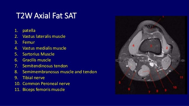
12 photos of the knee muscle anatomy mri. Click on the links to show each structure. Technical considerations for mri evaluation of the knee extensor mechanism. Atlas of anatomy in medical imagery. An understanding of normal anatomy and biomechanics of the knee extensor mechanism is necessary to comprehend the imaging of extensor mechanism injuries. Musculoskeletal radiology south texas radiology group. Want to learn more about it? Involved early gray = muscle: See the pictures and anatomy description of knee joint bones, cartilage, ligaments, muscle and tendons with resources for knee problems & injuries. Seems like it should be pretty easy, right? Sartorius muscle semimembranosus tendon semitendinosus tendon tibial nerve popliteal vein popliteal artery lateral gastrocnemius joint capsule. Anatomy of the knee can be complicated and hard to understand. Tips to keep joints healthy.
Injuries of the patellofemoral joint. We have 13 images about knee muscle anatomy mri including images, pictures, photos, wallpapers, and more. Articular surface of patella and femur, condyle, epicondyle and muscles (popliteus anatomy of the ankle and foot in mri: Want to learn more about it? On anatomical parts the user.
MRI KNEE JOINT ANATOMY from image.slidesharecdn.com Magnetic resonance imaging (mri scan): 12 photos of the knee muscle anatomy mri. Learn about knee anatomy muscle with free interactive flashcards. Use the checklist to quiz yourself. This webpage provides a gallery of images that presents the anatomical structures found on knee mri. Song, uc san francisco msiv gillian lieberman md. Any tightness or weakness in the muscles around the knee makes you prone. Click on the links to show each structure.
An understanding of normal anatomy and biomechanics of the knee extensor mechanism is necessary to comprehend the imaging of extensor mechanism injuries.
Musculoskeletal radiology south texas radiology group. Knee muscles need to have both good strength and flexibility. Contraction of the quadriceps group extends the leg. Want to learn more about it? An understanding of normal anatomy and biomechanics of the knee extensor mechanism is necessary to comprehend the imaging of extensor mechanism injuries. Master leg and knee anatomy using our topic page. Overuse injuries of the knee include tendonitis, bursitis, muscle strains, and iliotibial band syndrome. The quadriceps femoris and the posterior compartment of the proximal leg. Tips to keep joints healthy. Click on the links to show each structure. Knowing about knee anatomy can help people understand how knee arthritis develops and sometimes causes pain. Along the posterior portion of the muscle (yellow arrows), there is a flat area of tendon originating from the knee. This mri knee cross sectional anatomy tool is absolutely free to use.
Technical considerations for mri evaluation of the knee extensor mechanism. Use the checklist to quiz yourself. Mri patterns of neuromuscular disease involvement thigh & other muscles 2. If the knee is flexed more than 5 degrees, it may appear lax. Musculoskeletal radiology south texas radiology group.
How to Read Knee MRI of Normal Knee | Anatomy of the Knee | Colorado Knee Specialist - YouTube from i.ytimg.com In these page, we also have variety not only knee muscle anatomy mri, you could also find another pics such as axial knee mri, sagittal knee mri, mri axial knee anatomy, coronal. 12 photos of the knee muscle anatomy mri. The main knee muscles are the quadriceps, hamstrings and calf muscles. Mri knee anatomy cross patella sectional muscles sartorius femur surface epicondyle popliteus gastrocnemius muscle condyle atlas imaging body fascia. 1 november 2002 mri anatomy of the knee and shoulder james y. On anatomical parts the user. Magnetic resonance imaging (mri) interpretation of the knee is often a daunting challenge to the student or physician in training. Click now to learn more about the bones, muscles, and soft tissues of these regions at leg and knee anatomy:
Knee anatomy is incredibly complex, and problems with any part of the knee anatomy—including the bones, cartilage, muscles, ligaments and tendons—can cause pain.
Seems like it should be pretty easy, right? Song, uc san francisco msiv gillian lieberman md. Master leg and knee anatomy using our topic page. Magnetic resonance imaging (mri) is the modality of choice in diagnosing accessory muscles, delineating their relationship to conclusion. Articular surface of patella and femur, condyle, epicondyle and muscles (popliteus anatomy of the ankle and foot in mri: Find out about how the different muscles of the knee work and how they get injured. The hamstrings are a group of 3 muscles on the back of the thigh that provide the opposite motion by bending the knee from a straightened position. View of the anatomical labels. The main knee muscles are the quadriceps, hamstrings and calf muscles. 1 november 2002 mri anatomy of the knee and shoulder james y. Atlas of anatomy in medical imagery. Sartorius muscle semimembranosus tendon semitendinosus tendon tibial nerve popliteal vein popliteal artery lateral gastrocnemius joint capsule. Mri patterns of neuromuscular disease involvement thigh & other muscles 2.
Share :
Post a Comment
for "Knee Muscle Anatomy Mri / mri knee anatomy | knee sagittal anatomy | free cross sectional anatomy"


Post a Comment for "Knee Muscle Anatomy Mri / mri knee anatomy | knee sagittal anatomy | free cross sectional anatomy"