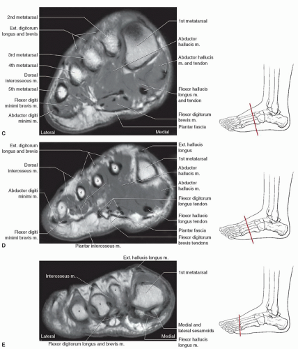
Foot Muscles Mri / Https Encrypted Tbn0 Gstatic Com Images Q Tbn And9gctles3hqgjfo8 Y77fwsabjyy3rtuvfbyzz0zt3gi4gooqavatv Usqp Cau. Indications for foot mri scan. The muscles of the neck can be divided into groups according to their location. The flexor digitorum brevis muscle lies immediately superior to the plantar aponeurosis and inferior to the tendons of the flexor digitorum longus in the sole of the foot. Magnetic resonance imaging (mri) is the modality of choice in diagnosing accessory muscles, delineating their relationship to adjacent structures, and differentiating them from soft tissue tumors. Accessory muscles are isointense to skeletal muscle on all pulse sequences, and can insert by fleshy muscular or tendinous insertions.
Coronal images are perpendicular to the long axis of the metatarsals. Foot muscles mri the extrinsic muscles are located in the anterior and lateral compartments of the leg. Electromyography in cases of foot drop involves testing of the tibialis anterior as well as muscles innervated by the superficial peroneal, tibial, sciatic, and superior gluteal nerves. Mri is the choice of modality for further imaging the ankle and foot after obtaining initial radiographs. This imaging technique assesses the ligaments and tendons, neurovascular structures (tarsal tunnel and plantar fascia), and the osseous structures(19).
Disease Activity Evident On Foot Mri During Clinical Remission In Rheumatoid Arthritis Rheumatology Advisor from www.rheumatologyadvisor.com They are mainly responsible for assisting some of the extrinsic muscles in their actions. Magnetic resonance imaging, otherwise known as mri, uses a combination of magnetic fields and radio waves to take images of the internal structures of your body. Adductor hallucis is anatomically located in the central compartment of foot, but the muscle is functionally grouped with the medial plantar muscles of foot because it acts on the great toe (hallux). Mri is an ideal method for identifying areas of muscle atrophy and fatty infiltration. Freitasrad is for educational purposes only and should not be used for medical treatment. The adductor hallucis has two heads: The aim of this review is to provide the reader with a comprehensive overview of the magnetic resonance imaging (mri) characteristics of the most common benign and malignant soft tissue neoplasms which occur around the foot and ankle. Indications for foot mri scan.
Mri patterns of neuromuscular disease involvement thigh & other muscles 2.
Distal part of the lateral and superior surfaces of the calcaneus and the apex of the inferior extensor retinaculum. Plantar interossei (foot) dr yuranga weerakkody ◉ and dr geon oh et al. In addition, an image of all the muscles of the back and plantar part of the foot, all tendons and tendon ligaments, blood vessels and nerves are obtained. Freitasrad is for educational purposes only and should not be used for medical treatment. Adductor hallucis is anatomically located in the central compartment of foot, but the muscle is functionally grouped with the medial plantar muscles of foot because it acts on the great toe (hallux). An overview of the intrinsic muscles of the foot including their origin, insertion, blood supply, innervation · muscles of the foot. The three plantar interossei muscles adduct the 3 rd, 4 th and 5 th toes toward the long axis through the 2 nd toe. The lower extremity mri for the foot and ankle is specifically designed to diagnose the following conditions: However, the roles of the extrinsic foot muscles during running have not been adequately identified. Mri is an ideal method for identifying areas of muscle atrophy and fatty infiltration. Coronal images are perpendicular to the long axis of the metatarsals. Mri with user outlined plantar intrinsic and extrinsic muscles group. Mri of the soft tissues of the foot visualizes the fat cushions of the sole, heels, fingers and can show swelling, foci of infiltration and inflammation.
Also known as osteomyelitis, which is generally treated with antibiotics, but can lead to an amputation. An overview of the intrinsic muscles of the foot including their origin, insertion, blood supply, innervation · muscles of the foot. • muscle edema is seen secondary to multiple etiologies including trauma, infectious and inflammatory processes, autoimmune disorders, neoplasms, and denervation injuries • on mri muscle edema is characterized by increase in free water within the muscle • muscle edema is seen on mri as increased signal on fluid sensitive sequences t2 fs Coronal images are perpendicular to the long axis of the metatarsals. Muscle anatomy knee mri 12 photos of the muscle anatomy knee mri muscle anatomy knee mri, human muscles, muscle anatomy knee mri
Wbct Mri Study Sheds Light On Flat Foot Degeneration Curvebeam from curvebeam.com The lower extremity mri for the foot and ankle is specifically designed to diagnose the following conditions: Magnetic resonance imaging (mri) is the modality of choice in diagnosing accessory muscles, delineating their relationship to adjacent structures, and differentiating them from soft tissue tumors. Your doctor, with the help of a radiologist, can then examine these images to determine whether there is anything wrong with your foot or ankle. Anatomy of the whole human body : Related posts of foot muscle anatomy mri muscle anatomy knee mri. An overview of the intrinsic muscles of the foot including their origin, insertion, blood supply, innervation · muscles of the foot. The purpose of this study was to examine the muscle functional (mf) mri and emg responses to perturbations of the foot by running in varus, neutral and valgus wedged shoes. Indications for foot mri scan.
Adductor hallucis is anatomically located in the central compartment of foot, but the muscle is functionally grouped with the medial plantar muscles of foot because it acts on the great toe (hallux).
• muscle edema is seen secondary to multiple etiologies including trauma, infectious and inflammatory processes, autoimmune disorders, neoplasms, and denervation injuries • on mri muscle edema is characterized by increase in free water within the muscle • muscle edema is seen on mri as increased signal on fluid sensitive sequences t2 fs The interosseous muscles of the foot are muscles found near the metatarsal bones that help to control the toes. Mri with user outlined plantar intrinsic and extrinsic muscles group. The aim of this review is to provide the reader with a comprehensive overview of the magnetic resonance imaging (mri) characteristics of the most common benign and malignant soft tissue neoplasms which occur around the foot and ankle. The flexor digitorum brevis muscle lies immediately superior to the plantar aponeurosis and inferior to the tendons of the flexor digitorum longus in the sole of the foot. Indications for foot mri scan. Lateral and medial processes of calcaneal tuberosity. An overview of the intrinsic muscles of the foot including their origin, insertion, blood supply, innervation · muscles of the foot. The purpose of this study was to examine the muscle functional (mf) mri and emg responses to perturbations of the foot by running in varus, neutral and valgus wedged shoes. Swelling and tenderness in your joints. Adductor hallucis is anatomically located in the central compartment of foot, but the muscle is functionally grouped with the medial plantar muscles of foot because it acts on the great toe (hallux). Freitasrad is for educational purposes only and should not be used for medical treatment. Coronal images are perpendicular to the long axis of the metatarsals.
• muscle edema is seen secondary to multiple etiologies including trauma, infectious and inflammatory processes, autoimmune disorders, neoplasms, and denervation injuries • on mri muscle edema is characterized by increase in free water within the muscle • muscle edema is seen on mri as increased signal on fluid sensitive sequences t2 fs The majority of soft tissue lesions in the foot and ankle are benign. Indications for foot mri scan. The lower extremity mri for the foot and ankle is specifically designed to diagnose the following conditions: Coronal images are perpendicular to the long axis of the metatarsals.
Foot Ankle And Calf Musculoskeletal Key from musculoskeletalkey.com Routine ankle magnetic resonance imaging (mri) tests involve taking images of the foot and ankle in the axial, coronal, and sagittal planes parallel to the tabletop(2). Magnetic resonance imaging, otherwise known as mri, uses a combination of magnetic fields and radio waves to take images of the internal structures of your body. Indications for foot mri scan. The interosseous muscles of the foot are muscles found near the metatarsal bones that help to control the toes. Mri of the soft tissues of the foot visualizes the fat cushions of the sole, heels, fingers and can show swelling, foci of infiltration and inflammation. Adductor hallucis is anatomically located in the central compartment of foot, but the muscle is functionally grouped with the medial plantar muscles of foot because it acts on the great toe (hallux). Its main symptoms include joint pain along with stiffness. Foot and (from schuenke m, schulte e.
Swelling and tenderness in your joints.
Mri with user outlined plantar intrinsic and extrinsic muscles group. Foot and (from schuenke m, schulte e. They are mainly responsible for assisting some of the extrinsic muscles in their actions. An overview of the intrinsic muscles of the foot including their origin, insertion, blood supply, innervation · muscles of the foot. General anatomy and the musculoskeletal system: Mri of the soft tissues of the foot visualizes the fat cushions of the sole, heels, fingers and can show swelling, foci of infiltration and inflammation. The muscles of the neck can be divided into groups according to their location. The flexor digitorum brevis muscle lies immediately superior to the plantar aponeurosis and inferior to the tendons of the flexor digitorum longus in the sole of the foot. The paraspinal muscles, which are innervated by the spinal nerve dorsal ramus, are also frequently tested. Mri is the choice of modality for further imaging the ankle and foot after obtaining initial radiographs. The interosseous muscles of the foot are muscles found near the metatarsal bones that help to control the toes. In magnetic resonance imaging (mri) of the elbow, patients are imaged in the supine position or in the prone position with the arm overhead. However, the roles of the extrinsic foot muscles during running have not been adequately identified.
Share :
Post a Comment
for "Foot Muscles Mri / Https Encrypted Tbn0 Gstatic Com Images Q Tbn And9gctles3hqgjfo8 Y77fwsabjyy3rtuvfbyzz0zt3gi4gooqavatv Usqp Cau"


Post a Comment for "Foot Muscles Mri / Https Encrypted Tbn0 Gstatic Com Images Q Tbn And9gctles3hqgjfo8 Y77fwsabjyy3rtuvfbyzz0zt3gi4gooqavatv Usqp Cau"